The Role of Advanced Airways in ACLS
When Airway Management Becomes a Priority
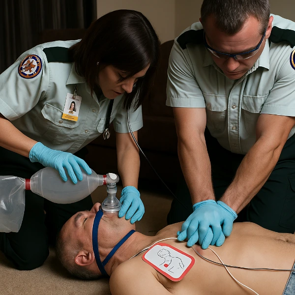
Effective airway management during cardiac arrest can improve patient outcomes, but only when timed appropriately. High-quality compressions and early defibrillation remain the foundation of advanced cardiovascular life support (ACLS). An advanced airway should only be introduced when it enhances oxygenation without causing delays. Rescuers must assess for ineffective bag-valve-mask (BVM) ventilation or signs of airway compromise. Those moments justify escalation to a supraglottic airway or endotracheal tube. Any intervention must fit within the broader ACLS framework without disrupting circulation.
Devices Used: BVM vs Supraglottic vs ETT
Selecting the right airway device depends on provider skill, patient anatomy, and available equipment. BVM remains the initial tool of choice and works well when two providers maintain a good seal. Supraglottic airways, like the i-gel or laryngeal mask airway, are fast and effective when intubation is delayed or not possible. Endotracheal intubation offers definitive control but requires training and often momentary interruption of compressions. Decision-making should weigh airway protection, ventilation efficiency, and team capabilities.
Rapid Sequence Intubation (RSI): What It Is and Why It Matters
Defining RSI for Emergency Cardiac Care
Rapid sequence intubation (RSI) involves giving a sedative followed quickly by a paralytic to optimize conditions for intubation. This approach minimizes gag reflexes and muscle tone, allowing smoother tube placement. ACLS teams use RSI when a patient is unconscious but not in full arrest, or when post-arrest ventilation needs escalate. RSI helps prevent aspiration and improves first-pass success rates. Teams must assess whether the patient can tolerate apnea and whether benefits outweigh risks.

Drug Protocols for RSI in the Field
Drug choice matters, especially in prehospital settings. Etomidate is a common induction agent due to its cardiovascular stability. Ketamine offers an alternative, especially for hypotensive patients, because it supports blood pressure. For paralytics, succinylcholine acts fast but can cause complications like hyperkalemia. Rocuronium provides a longer duration with fewer contraindications. Crews must also consider drug compatibility, dosing accuracy, and timing—especially when transitioning from spontaneous breathing to controlled ventilation. RSI requires careful planning and skilled execution.
RSI and Cardiac Arrest Medications: Working Together
ACLS medications such as epinephrine and amiodarone may be administered around the same time as RSI. Coordinating timing is crucial to avoid overlapping adverse effects. For instance, epinephrine-induced tachycardia may exaggerate ketamine’s sympathomimetic effects. Amiodarone may prolong QT intervals, compounding risks with paralytics. Teams must communicate precisely about what has been given and when. This coordination ensures both drug safety and procedural success.
Preparing for the Airway: Preoxygenation, Positioning & Backups
How to Maximize Apneic Time Safely
Preoxygenation extends the safe window for intubation by filling the lungs with oxygen. Using a non-rebreather mask or BVM with high-flow oxygen helps maximize reserves. Adding nasal cannula oxygen during the procedure supports apneic oxygenation. Patients should be positioned with their head elevated or in a ramped configuration. These adjustments enhance diaphragmatic excursion and delay desaturation. Field providers should practice these maneuvers regularly to improve performance under pressure.
Have a Backup Plan — Always
Even skilled intubators face failed attempts. Supraglottic airways should be ready as a backup for difficult airways or failed intubation. Bag-mask ventilation may still be the safest option if insertion delays compressions. Providers should know when to stop and pivot. If multiple attempts fail or oxygen saturation drops quickly, rescuers must consider emergency cricothyrotomy. Training, teamwork, and rehearsal of these algorithms can improve success during actual emergencies.
Equipment Redundancy and Checklists
Carrying multiple airway tools is critical. Teams should pack redundant suction devices, back-up BVMs, multiple airway adjuncts, and checklists for RSI. These visual tools help ensure nothing gets missed during high-stress situations. An overlooked stylet or malfunctioning syringe can stall the entire intubation. Checklists and preparation drills can prevent small oversights from becoming critical errors. Reliable airway access begins with smart packing.
Airway Placement: Techniques and Confirmation
Laryngoscopy Choices and First-Pass Success
First-pass success improves outcomes and reduces hypoxia risk. Direct laryngoscopy (DL) remains widely used, but video laryngoscopy (VL) continues to gain ground. VL offers a better glottic view and improves intubation in difficult anatomy. Providers should choose the method they are most comfortable with, but should also train in both. Using a bougie or stylet helps guide the tube through the vocal cords. Positioning and suction readiness also contribute to successful placement.
Confirming Tube Placement During Resuscitation
Visual confirmation of tube passing the cords is not enough. Quantitative waveform capnography provides reliable feedback that confirms endotracheal placement. This technology measures exhaled CO₂ and displays a waveform, making it invaluable during CPR. ETCO₂ should rise as circulation improves, giving clues about perfusion quality. Chest rise and bilateral breath sounds can supplement but should never replace waveform confirmation. In cardiac arrest, monitoring capnography trends also guides return of spontaneous circulation.
When to Reassess or Replace the Airway
Not all intubations remain successful. A misplaced tube or dislodgment can cause hypoxia. Providers must continuously monitor chest rise, SpO₂, and capnography trends. If waveform disappears or saturation drops rapidly, reassessment is mandatory. Tube displacement may occur during movement, especially in chaotic or confined scenes. A supraglottic device may need to be placed if repeated attempts delay oxygenation. Timely reassessment prevents prolonged hypoxia and preserves neurologic outcomes.
Avoiding Pitfalls in Cardiac Arrest Intubation
Compression Interruptions & Survival Risk
Every second without chest compressions reduces coronary and cerebral perfusion. Studies show that prolonged intubation attempts can lower survival rates. Teams must coordinate to intubate during rhythm checks or short pauses. Experienced providers may even attempt intubation during compressions when feasible. Simulation training helps crews practice minimizing hands-off time. The goal is to secure the airway with minimal delay, not to achieve a textbook-perfect tube placement.
Preventing Desaturation, Hypotension & Arrest
Preoxygenation and ongoing oxygen delivery reduce desaturation risks. Some patients may benefit from fluid boluses or vasopressors before RSI to reduce the risk of post-intubation hypotension. Etomidate and ketamine remain the safest induction agents in these cases. Teams must observe for sudden changes in blood pressure or cardiac rhythm. If needed, adjust ventilation rates to avoid hyperventilation. Post-intubation sedation also prevents agitation and maintains tube security. These steps preserve patient stability in fragile moments.
Chart: Common Complications and Countermeasures
| Pitfall | Underlying Cause | Suggested Countermeasure |
|---|---|---|
| Prolonged intubation attempts | Inexperienced operator or poor visibility | Switch to supraglottic airway after 2 failed attempts |
| Desaturation during laryngoscopy | Inadequate preoxygenation or long apnea | Use nasal cannula with NRB or BVM during intubation |
| Post-RSI hypotension | Drug-induced vasodilation or cardiac depression | Use ketamine; administer fluid bolus beforehand |
| Tube displacement in transport | Unsecured ETT or patient movement | Recheck placement often; tape or secure tube well |
What EMS Clinicians Should Practice and Document
Training for Real-World ACLS Scenarios
EMS crews must regularly practice RSI and airway management through hands-on stations. These drills should simulate real cardiac arrests with compressed timelines and dynamic roles. Skills like assembling equipment, calculating drug doses, and confirming placement must become second nature. Teams should rehearse failed airway drills and switch roles between sessions. Incorporating scenario-based evaluation improves confidence and performance during actual ACLS calls.
Documentation Essentials: Time, Tools & Tube Placement
Accurate records support quality improvement and legal protection. Providers must document the time of intubation, drugs administered, and confirmation methods. ETCO₂ values, breath sounds, and chest rise observations also belong in the record. Teams should include details on preoxygenation strategy and complications encountered. Digital systems may prompt this input, but paper forms must capture it too. Clear documentation also supports post-event debriefs and performance reviews.
Essential Takeaways for Airway Success in ACLS
Clear, timely airway management can save lives—but only when executed with skill and precision. RSI offers better control but demands a well-coordinated team and a confident operator. Crews must prioritize oxygenation, limit pauses in compressions, and confirm placement with capnography. Supraglottic airways remain valuable backups, not last resorts. Training frequently, documenting completely, and reviewing cases help improve performance with every shift.
For readers planning to enhance their clinical skills or pursue ACLS certification, the ACLS course page at EMS Ricky offers structured training with live scenarios. If you’re unsure how to get started, our application process guide walks you through every step.
FAQ: Airway and RSI in ACLS
What is the ideal timing for intubation during cardiac arrest?
Intubation should not delay high-quality CPR or defibrillation. It’s best performed during rhythm checks or when bag-mask ventilation fails. Teams must weigh the need for an advanced airway against the risk of interrupting chest compressions. Delayed or poorly timed intubation may decrease survival. Supraglottic devices may offer a safer interim solution.
Which RSI drug combination is preferred in hypotensive patients?
Ketamine paired with rocuronium is typically preferred. Ketamine supports blood pressure through its sympathomimetic effects, while rocuronium has fewer contraindications than succinylcholine. Etomidate may still be used for its hemodynamic neutrality. The goal is to avoid drops in perfusion that could worsen brain injury or delay recovery.
How can crews confirm tube placement in chaotic environments?
Waveform capnography is the most reliable tool. It confirms carbon dioxide exchange, even during CPR. Crews should also observe chest rise and listen for bilateral breath sounds, but these signs may be misleading. Frequent rechecks ensure the tube remains in place, especially during transport or patient movement.
Why is video laryngoscopy gaining popularity in EMS?
Video laryngoscopy improves visualization of the vocal cords, especially in difficult airways. It allows providers to see around anatomical obstructions and confirm placement with better accuracy. The shared view also facilitates training and real-time team coaching. Still, it requires practice and may fail in blood or secretion-filled fields.
Should RSI be attempted in all cardiac arrest patients?
No. In patients who are pulseless and unresponsive, simple BVM ventilation or an SGA may suffice. RSI is typically reserved for patients with a gag reflex, post-ROSC ventilation, or severe airway compromise. The decision should be clinical, team-based, and aligned with ACLS goals—not automatic or routine.
